جمعه ۲۴ شهریور ۹۶ ۱۵:۱۷ ۱۶ بازديد
Overview
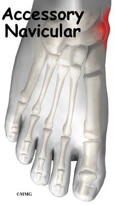
An accessory navicular bone is an accessory bone of the foot that occasionally develops abnormally causing a plantar medial enlargement of the navicular. The accssory navicular bone presents as a sesamoid in the posterior tibial tendon, in articulation with the navicular or as an enlargment of the navicular. Navicular (boat shaped) is an intermediate tarsal bone on the medial side of the foot. It is located on the medial side of the foot, and articulates proximally with the talus. Distally it articulates with the three cuneiform bones. In some cases it articulates laterally with the cuboid. The tibialis posterior inserts to the os naviculare. The tibialis posterior muscle also contracts to produce inversion of the foot and assists in the plantar flexion of the foot at the ankle. Tibialis posterior also has a major role in supporting the medial arch of the foot. This supports is compromised by abnormal insertion of the tendon into the accessory navicular bone when present. This lead to loss of suspension of tibialis posterior tendon and may cause peroneal spastic pes planus or simple pes planus. But, yet a cause and effect relationship between the accessory navicular and pes planus is doubtful and is yet unproved clearly.

Causes
It is commonly believed that the posterior tibial tendon loses its vector of pull to heighten the arch. As the posterior muscle contracts, the tendon is no longer pulling straight up on the navicular but must course around the prominence of bone and first pull medially before pulling upward. In addition, the enlarged bones may irritate and damage the insertional area of the posterior tibial tendon, making it less functional. Therefore, the presence of the accessory navicular bone does contribute to posterior tibial dysfunction.
Symptoms
Adolescence is a common time for the symptoms to first appear. This is a time when bones are maturing and cartilage is developing into bone. Sometimes, however, the symptoms do not occur until adulthood. The signs and symptoms of accessory navicular syndrome include a visible bony prominence on the midfoot (the inner side of the foot, just above the arch) Redness and swelling of the bony prominence, Vague pain or throbbing in the midfoot and arch, usually occurring during or after periods of activity.
Diagnosis
Your podiatrist will most likely diagnose accessory navicular syndrome by making a visual study of the area, checking whether the shape of your foot and your ability to move it indicate there?s an accessory navicular lurking around. He or she may push on the prominence on your foot to check to see if it hurts, and may ask you to walk around in order to ascertain How can we increase our height? your gait is affected. In order to get a certain diagnosis, your podiatrist will need some way to see the inside of your foot, which will most likely involve getting X-rays, or possibly an MRI or some other scan of your foot?s interior.
Non Surgical Treatment
Excess weight will increase the force on the posterior tibial tendon as it inserts into the accessory navicular and will tend to precipitate or aggravate symptoms. If a patient with a symptomatic accessory navicular is overweight, then losing weight can be very helpful. Even losing 5-10lbs will decrease the amount of force going through the foot with each step by as much as 15-30lbs. This is because the foot acts like a lever serving to magnify the force absorbed by the foot with each step.
Surgical Treatment
The original procedure advocated by Kidner involved shelling out of the accessory navicular bone from within the insertional area of the posterior tibial tendon and rerouting this tendon under the navicular bone in hopes of restoring a normal pull of this tendon. When treating younger children, history has shown us that simply shelling out of the accessory navicular bone from within the tendon and remodeling the tuberosity of the navicular bone can give you satisfactory results.
In general, you want to reserve advancement of the posterior tibial tendon for adults or those who have a more significant flatfoot deformity. You may also use this approach after determining that quality custom orthotics are only resulting in a slight decrease of symptoms.
Th1s1sanart1cl3s1te
An accessory navicular bone is an accessory bone of the foot that occasionally develops abnormally causing a plantar medial enlargement of the navicular. The accssory navicular bone presents as a sesamoid in the posterior tibial tendon, in articulation with the navicular or as an enlargment of the navicular. Navicular (boat shaped) is an intermediate tarsal bone on the medial side of the foot. It is located on the medial side of the foot, and articulates proximally with the talus. Distally it articulates with the three cuneiform bones. In some cases it articulates laterally with the cuboid. The tibialis posterior inserts to the os naviculare. The tibialis posterior muscle also contracts to produce inversion of the foot and assists in the plantar flexion of the foot at the ankle. Tibialis posterior also has a major role in supporting the medial arch of the foot. This supports is compromised by abnormal insertion of the tendon into the accessory navicular bone when present. This lead to loss of suspension of tibialis posterior tendon and may cause peroneal spastic pes planus or simple pes planus. But, yet a cause and effect relationship between the accessory navicular and pes planus is doubtful and is yet unproved clearly.
Causes
It is commonly believed that the posterior tibial tendon loses its vector of pull to heighten the arch. As the posterior muscle contracts, the tendon is no longer pulling straight up on the navicular but must course around the prominence of bone and first pull medially before pulling upward. In addition, the enlarged bones may irritate and damage the insertional area of the posterior tibial tendon, making it less functional. Therefore, the presence of the accessory navicular bone does contribute to posterior tibial dysfunction.
Symptoms
Adolescence is a common time for the symptoms to first appear. This is a time when bones are maturing and cartilage is developing into bone. Sometimes, however, the symptoms do not occur until adulthood. The signs and symptoms of accessory navicular syndrome include a visible bony prominence on the midfoot (the inner side of the foot, just above the arch) Redness and swelling of the bony prominence, Vague pain or throbbing in the midfoot and arch, usually occurring during or after periods of activity.
Diagnosis
Your podiatrist will most likely diagnose accessory navicular syndrome by making a visual study of the area, checking whether the shape of your foot and your ability to move it indicate there?s an accessory navicular lurking around. He or she may push on the prominence on your foot to check to see if it hurts, and may ask you to walk around in order to ascertain How can we increase our height? your gait is affected. In order to get a certain diagnosis, your podiatrist will need some way to see the inside of your foot, which will most likely involve getting X-rays, or possibly an MRI or some other scan of your foot?s interior.
Non Surgical Treatment
Excess weight will increase the force on the posterior tibial tendon as it inserts into the accessory navicular and will tend to precipitate or aggravate symptoms. If a patient with a symptomatic accessory navicular is overweight, then losing weight can be very helpful. Even losing 5-10lbs will decrease the amount of force going through the foot with each step by as much as 15-30lbs. This is because the foot acts like a lever serving to magnify the force absorbed by the foot with each step.
Surgical Treatment
The original procedure advocated by Kidner involved shelling out of the accessory navicular bone from within the insertional area of the posterior tibial tendon and rerouting this tendon under the navicular bone in hopes of restoring a normal pull of this tendon. When treating younger children, history has shown us that simply shelling out of the accessory navicular bone from within the tendon and remodeling the tuberosity of the navicular bone can give you satisfactory results.
In general, you want to reserve advancement of the posterior tibial tendon for adults or those who have a more significant flatfoot deformity. You may also use this approach after determining that quality custom orthotics are only resulting in a slight decrease of symptoms.
Th1s1sanart1cl3s1te